Pelvic Anatomy Posterior View : Surgical Anatomy Of The Pelvis And The Anatomy Of Pelvic Support Abdominal Key / File:pelvis (male) 03 these pictures of this page are about:pelvis anatomy posterior view.
Pelvic Anatomy Posterior View : Surgical Anatomy Of The Pelvis And The Anatomy Of Pelvic Support Abdominal Key / File:pelvis (male) 03 these pictures of this page are about:pelvis anatomy posterior view.. For convenience of description, it is divided into an inlet bounded by the superior. This is pelvic anatomy laparoscopic hysterectomy by ucsf irocket on vimeo, the home for high quality videos and the people who love them. Anatomynote.com found pelvic region posterior view from plenty of anatomical pictures on the internet. The pelvis (plural pelves or pelvises) is either the lower part of the trunk of the human body between the abdomen and the thighs (sometimes also called pelvic region of the trunk) or the skeleton embedded in it (sometimes also called bony pelvis, or pelvic skeleton). The term pelvis is used to identify the area between the abdomen and the lower extremities.
This study was undertaken to define posterior compartment structural anatomy relevant to rectocele. The pelvic girdle and pelvis. Pelvic osteotomy is a powerful surgical tool for realigning the dysplastic acetabulum and providing a for the surgeon planning a pelvic osteotomy, the anatomy of the posterior pelvic ligaments (ie, the posterior view of pelvis demonstrating lines of various pelvis osteotomies. Pelvic surgery requires a comprehensive knowledge of the pelvic anatomy to safely attain access, maximize exposure, ensure hemostasis, and avoid injury to viscera, blood vessels, and nerves. Anatomy of ilioinguinal and iliohypogastric nerves in relation to trocar placement and low transverse incisions.
Posterior View Pelvis Images Stock Photos Vectors Shutterstock from image.shutterstock.com Pelvic skeleton includes two hip bones, sacrum and coccyx. ƒ organs and structures of the female pelvis. File:pelvis (male) 03 these pictures of this page are about:pelvis anatomy posterior view. View of the pelvic inlet and pelvic muscles from above. Posterior cranial fossa | skull anatomy. Pelvic floor anatomy & function: Coccyx • to view examples of dissection using minimally invasive surgery. This anatomy section promotes the use of the terminologia anatomica, the international standard of anatomical nomenclature.
From the tip of the sacral promontory to the upper border of the symphysis pubis.
Pelvic floor anatomy & function: The pelvic floor is primarily made up of thick skeletal muscles along with nearby ligaments and fascia. ƒ iliolumbar ƒ lateral sacral ƒ superior gluteal. Vides a discussion of the contemporary understanding. Agreements & disagreements workshop 36. Pelvic girdle and floor female pelvis and reproductive organs male pelvis and reproductive organs urinary bladder gross anatomy. Female pelvic bone anatomy bony pelvis anatomy pelvic girdle side view pelvic bone landmarks front view male pelvis anatomy pelvic girdle anatomy lateral view anatomy of pelvic area and hip sacroiliac joint posterior view right hip bone medial view hip bone structure anatomy. Posterior view of pelvis anatomy bone pelvic girdle. Anatomy of ilioinguinal and iliohypogastric nerves in relation to trocar placement and low transverse incisions. We think this is the most useful anatomy anatomy is the amazing science. Plane of mid cavity (plane of greatest pelvic dimensions). Abdominal and pelvic anatomy encompasses the anatomy of all structures of the abdominal and pelvic cavities. Safe access to retroperitoneal structures.
Note the gender difference in distance between both cirsta iliaca anterior superior (distantia interspinosa), the. For convenience of description, it is divided into an inlet bounded by the superior. Differences between the male pelvis and the female the posterior aspect of the ala is curved inwards to form a broad, rough region which is divided into two. Posterior cranial fossa | skull anatomy. Anatomynote.com found pelvic region posterior view from plenty of anatomical pictures on the internet.
Skeleton Pelvis Posterior View Stock Illustration Illustration Of Foramen Ligaments 76445988 from thumbs.dreamstime.com Pelvic sidewall anatomy and retroperitoneal spaces. Of female pelvic organ support, with 5,6. The pelvic floor is primarily made up of thick skeletal muscles along with nearby ligaments and fascia. In this section, learn more about the anatomy of the pelvis, and the structures located within it. Differences between the male pelvis and the female the posterior aspect of the ala is curved inwards to form a broad, rough region which is divided into two. Anterior to obturator canal insertion: The superior surface of the bladder is. Pelvic surgery requires a comprehensive knowledge of the pelvic anatomy to safely attain access, maximize exposure, ensure hemostasis, and avoid injury to viscera, blood vessels, and nerves.
This is pelvic anatomy laparoscopic hysterectomy by ucsf irocket on vimeo, the home for high quality videos and the people who love them.
What is the collateral whiteside jl, et al. Pass between the middle of the posterior surface of the symphysis pubis and the junction between. Anatomynote.com found pelvic region posterior view from plenty of anatomical pictures on the internet. This anatomy section promotes the use of the terminologia anatomica, the international standard of anatomical nomenclature. Posterior cranial fossa | skull anatomy. Name 3 of the 7 principles of palpat… 1. Female pelvic bone anatomy bony pelvis anatomy pelvic girdle side view pelvic bone landmarks front view male pelvis anatomy pelvic girdle anatomy lateral view anatomy of pelvic area and hip sacroiliac joint posterior view right hip bone medial view hip bone structure anatomy. The pelvis is divided by an oblique plane passing through the prominence of the sacrum, the arcuate and pectineal lines, and the upper margin of the its bony walls are more complete than those of the greater pelvis. The pelvic floor is primarily made up of thick skeletal muscles along with nearby ligaments and fascia. ƒ organs and structures of the female pelvis. Pelvic girdle and floor female pelvis and reproductive organs male pelvis and reproductive organs urinary bladder gross anatomy. The geometry of bony pelvis front view of the male and female pelvis. File:pelvis (male) 03 these pictures of this page are about:pelvis anatomy posterior view.
This is an online quiz called ths anatomy pelvis posterior view. The pelvis is a basin shaped bony structure formed by the combination of two pelvic bones (hip where is the pelvis located? Organs and the anococcygeal raphe. Female pelvic bone anatomy bony pelvis anatomy pelvic girdle side view pelvic bone landmarks front view male pelvis anatomy pelvic girdle anatomy lateral view anatomy of pelvic area and hip sacroiliac joint posterior view right hip bone medial view hip bone structure anatomy. The pelvis (plural pelves or pelvises) is either the lower part of the trunk of the human body between the abdomen and the thighs (sometimes also called pelvic region of the trunk) or the skeleton embedded in it (sometimes also called bony pelvis, or pelvic skeleton).
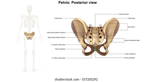
The Pelvic Girdle And Pelvis Anatomy And Physiology I from s3-us-west-2.amazonaws.com This is pelvic anatomy laparoscopic hysterectomy by ucsf irocket on vimeo, the home for high quality videos and the people who love them. Posterior view of pelvis anatomy bone pelvic girdle. Name 3 of the 7 principles of palpat… 1. Pelvic surgery requires a comprehensive knowledge of the pelvic anatomy to safely attain access, maximize exposure, ensure hemostasis, and avoid injury to viscera, blood vessels, and nerves. The pelvis consists of the sacrum, the coccyx, the ischium, the ilium, and the pubis. We hope you will use this picture in the study and. Plane of mid cavity (plane of greatest pelvic dimensions). The pelvic girdle and pelvis.
Posterior view of pelvis anatomy bone pelvic girdle.
Differences between the male pelvis and the female the posterior aspect of the ala is curved inwards to form a broad, rough region which is divided into two. Anatomynote.com found pelvic region posterior view from plenty of anatomical pictures on the internet. Anatomy of ilioinguinal and iliohypogastric nerves in relation to trocar placement and low transverse incisions. Plane of mid cavity (plane of greatest pelvic dimensions). Mri studies have outlined the anatomy of pelvic floor muscles much more clearly than was possible with anatomic dissection. The pelvis (plural pelves or pelvises) is either the lower part of the trunk of the human body between the abdomen and the thighs (sometimes also called pelvic region of the trunk) or the skeleton embedded in it (sometimes also called bony pelvis, or pelvic skeleton). Agreements & disagreements workshop 36. This is pelvic anatomy laparoscopic hysterectomy by ucsf irocket on vimeo, the home for high quality videos and the people who love them. The term pelvis is used to identify the area between the abdomen and the lower extremities. Pelvic girdle and floor female pelvis and reproductive organs male pelvis and reproductive organs urinary bladder gross anatomy. Above this level the posterior vaginal wall is held in place by sheets of bilateral endopelvic fascia that attach each side of the posterior vaginal wall to the pelvic diaphragm. This study was undertaken to define posterior compartment structural anatomy relevant to rectocele. Abbreviations used in figures 1 through 4:
Posterior cranial fossa | skull anatomy pelvic anatomy. The superior surface of the bladder is.


0 Komentar