Lower Body Bone Anatomy - Lower Limb Clinical Gate / Bones provide the primary support and structure for the body, but they also do much more.. (also name is it anterior or posterior? These sections are cervical (neck), thoracic (upper and middle back), lumbar (lower back), and sacrum (tailbone). Get product information, training, support, solutions and more. Its body forms 1/5 of the acetabulum; They provide a great deal of strength to modulate powerful forces between the upper and lower body.
This curve, called lordosis, helps to: The superior and inferior pubic rami participate in the formation of the obturator foramen Its symphyseal surface unites with the pubis of the opposite side to form the pubic symphysis; The femur is the single bone of the thigh. The tibia is the main bone of the lower leg, forming what is more commonly known as the shin.
Lower Limb Bones Muscles Joints Nerves How To Relief Anatomy Bones Medical Anatomy Human Skeleton Anatomy from i.pinimg.com (also name is it anterior or posterior? Its a little intense, oh and spelling counts! Anatomy of shoulder 12 photos of the anatomy of shoulder anatomy of nerves in shoulder, anatomy of posterior shoulder dislocation, anatomy of right shoulder, anatomy of shoulder labrum tear, anatomy of the shoulder games, human anatomy, anatomy of nerves in shoulder, anatomy of posterior shoulder dislocation, anatomy of right. The medial side of the tibia is located immediately under the skin, allowing it to be easily palpated down the entire length of the medial leg. It is divisible into three portions—a body and two rami. Lower extremity anatomy the lower extremity refers to the human leg, including the gluteal or hip region, thigh and foot. Muscles of the lower limb. Laterally it is thicker and the hairs are more numerous.
The thigh bone, or femur, is the large upper leg bone that connects the lower leg bones (knee joint) to the pelvic bone (hip joint).
All the bones in the body can be described as long bones or flat bones. The femur is the single bone of the thigh. Anatomy of shoulder 12 photos of the anatomy of shoulder anatomy of nerves in shoulder, anatomy of posterior shoulder dislocation, anatomy of right shoulder, anatomy of shoulder labrum tear, anatomy of the shoulder games, human anatomy, anatomy of nerves in shoulder, anatomy of posterior shoulder dislocation, anatomy of right. The osteology of the lower limb is particularly detailed, with 3d view and patterns of bone structures and muscle insertions and ligaments of the hip bone, the femur, the patella, tibia, the fibula, tibial plateau, the tibial pilon, the foot (talus, calcaneus, cuboid, cuneiform bones, metatarsal bones, phalanges proximal, middle and distal). Bones also house bone marrow, which helps to produce a number of blood cell types that are vital to healthy body function. They have a vital role in maintaining the body's mineral composition and protect vital organs from harm. Surface anatomy of the lower extremity. The back of the thigh from the sits bones to the top of the lower leg, crossing the knee joint. 1 your spine in this region has a natural inward curve. Laterally it is thicker and the hairs are more numerous. The lower extremity includes the hip, knee, and ankle joints, and the bones of the thigh, leg, and foot. Related posts of anatomy of lower body anatomy of shoulder. Get product information, training, support, solutions and more.
Bones provide the primary support and structure for the body, but they also do much more. Surface anatomy of the lower extremity. The study of the human body involves anatomy, physiology, histology and. —the ischium forms the lower and back part of the hip bone. Function of the hamstring muscles the main functions of the hamstrings are to flex (bend) the knee and extend the hip.
Lower Extremity Anatomy Bones Muscles Nerves Vessels Kenhub from thumbor.kenhub.com They have a vital role in maintaining the body's mineral composition and protect vital organs from harm. Learn how to communicate up to six times better by using visuals with smartdraw. The lower leg contains two major long bones, the tibia and the fibula, which are both very strong skeletal structures. The osteology of the lower limb is particularly detailed, with 3d view and patterns of bone structures and muscle insertions and ligaments of the hip bone, the femur, the patella, tibia, the fibula, tibial plateau, the tibial pilon, the foot (talus, calcaneus, cuboid, cuneiform bones, metatarsal bones, phalanges proximal, middle and distal). The tibia (also called the shinbone) is located near the midline of the leg. In fact, the leg is the part of the body between the knee and ankle joints. Many people refer to the lower extremity as the leg. The sacroiliac (si) joints connect the sacrum at the base of the spine with the hip bone.
Learn how to communicate up to six times better by using visuals with smartdraw.
Bones provide the primary support and structure for the body, but they also do much more. The proper way to describe the lower limb is the lower extremity. Articulating at the knee and ankle joints respectively. Related posts of anatomy of lower body anatomy of shoulder. They build the connection between the lower leg and the metatarsus. Pelvic girdle the pelvic girdle contains the hip bone and the sacrum. Each hip bone has three parts (ilium, ischium, pubis) and accepts the head of the femur to form the hip joint. Its body forms 1/5 of the acetabulum; (include if it is posterior or anterior and what side of the body it is on) 2. The back of the thigh from the sits bones to the top of the lower leg, crossing the knee joint. They provide a great deal of strength to modulate powerful forces between the upper and lower body. Nerves of the lower limb. Your lower back (lumbar spine) is the anatomic region between your lowest rib and the upper part of the buttock.
An angulated bone the forms the anterior part of the pelvis: The lower leg contains two major long bones, the tibia and the fibula, which are both very strong skeletal structures. Recall that the iliac crest contains the What side of the body is it on?) 3. Bones provide the primary support and structure for the body, but they also do much more.
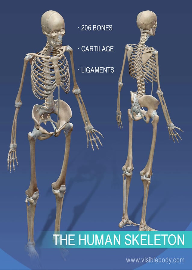
Overview Of Skeleton Learn Skeleton Anatomy from www.visiblebody.com One of three bones that form the os coxae: An angulated bone the forms the anterior part of the pelvis: The osteology of the lower limb is particularly detailed, with 3d view and patterns of bone structures and muscle insertions and ligaments of the hip bone, the femur, the patella, tibia, the fibula, tibial plateau, the tibial pilon, the foot (talus, calcaneus, cuboid, cuneiform bones, metatarsal bones, phalanges proximal, middle and distal). Recall that the iliac crest contains the Upgrade and get a lot more done! This part of the interactive atlas of anatomy of the human body is about the arterial vasculature of the pelvic girdle, pelvis, thigh, knee, leg and foot and the. It is divisible into three portions—a body and two rami. The tibia is the main bone of the lower leg, forming what is more commonly known as the shin.
One of three bones that form the os coxae:
It is composed of 300 bones at birth, but later decreases to 80 bones in the axial skeleton and 126 bones in the appendicular skeleton. The femur is the single bone of the thigh. Lower leg anatomy the lower leg extends from the knee to the ankle. The lower limb contains 30 bones. The study of the human body involves anatomy, physiology, histology and. Recall that the iliac crest contains the The back of the thigh from the sits bones to the top of the lower leg, crossing the knee joint. However, the exact definition of lower extremity in the human anatomy basically refers to that portion of your lower limb which extends from your knee till the ankle. The vertebral column of the lower back includes the five lumbar vertebrae, the sacrum, and the coccyx. The proper way to describe the lower limb is the lower extremity. Your lower back (lumbar spine) is the anatomic region between your lowest rib and the upper part of the buttock. (also name is it anterior or posterior? The skeletal system also provides attachment points for muscles to allow movements at the joints.
In fact, the leg is the part of the body between the knee and ankle joints anatomy lower body. Lower leg anatomy the lower leg extends from the knee to the ankle.


0 Komentar